
Free AI summary of this YouTube video by SumTube
How to Interpret a Chest X-Ray (Lesson 1 - An Introduction)
TL;DR
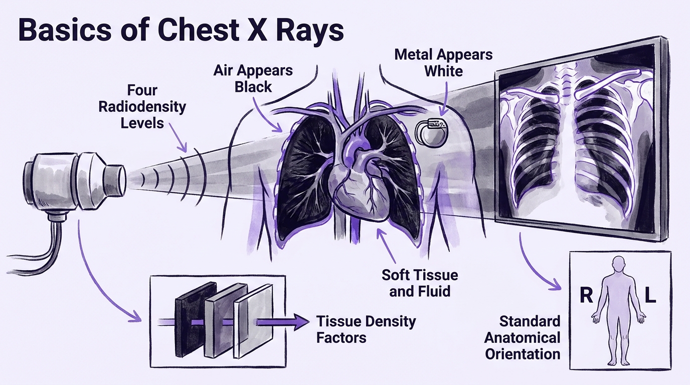
This introductory lesson explains the basic physics, equipment, and indications behind acquiring conventional chest X-rays. It covers how density, thickness, and exposure duration determine radiodensity across four main tissue classes (air, fluid/soft tissue, bone, and metal). Finally, it outlines the standard PA, lateral, and portable AP views while introducing the structure of the 10-lesson interpretation course.

Key takeaways
- 10:41X-ray film works in reverse of intuitive expectation: unexposed areas remain white, while fully exposed areas turn black
- 9:02Image brightness on a film is governed by three factors: physical tissue density/atomic mass, path thickness, and exposure duration
- 13:21Medical X-rays categorise body materials into four main radiodensity levels: air (radiolucent), soft tissue/fluid, bone, and metal (radiopaque)
- 19:58Standard chest X-rays are displayed in anatomical orientation, meaning the viewer's left corresponds to the patient's right side
Tap to enlarge
Most valuable section
For whom
Medical students, nurses, and clinicians seeking a clear conceptual foundation of X-ray physics and tissue radiodensity.
Physics of X-Ray Density and Exposure9:02
It uses clear glass-jar analogies to demonstrate exactly how tissue thickness, density, and exposure time manipulate shades of gray on film.
Detailed summary
1. Course Overview and Learning Objectives
0:12This section introduces a 10-lesson course designed to teach non-radiologist healthcare professionals how to systematically interpret chest X-rays. The curriculum spans basic X-ray physics, film quality assessment, systematic anatomical review, common pathologies, line and tube placement, and a self-assessment clinical vignette case review.
The primary learning objectives for this first video include understanding the basic physics behind X-ray generation, learning how shadows form on film, and recognizing the differences between standard chest X-ray projections such as PA, lateral, and AP views.
2. Clinical Indications for Chest X-Rays
3:41Chest X-rays are among the most common non-invasive diagnostic tests in clinical medicine. Standard indications include evaluating respiratory or cardiac symptoms (cough, shortness of breath, chest pain, fever), assessing physical exam findings like hypoxemia, verifying line or tube placement, and screening for pneumothorax following invasive procedures.
Conversely, routine pre-operative chest X-rays for asymptomatic patients are inappropriate and offer no clinical benefit. Similarly, standard chest X-rays should not be used as routine lung cancer screening tools in smokers, where CT imaging is preferred.
3. Physics of X-Ray Density and Exposure
5:40X-rays are high-energy photons capable of penetrating body tissues, but they also carry a risk of DNA damage that requires limiting exposure. An X-ray setup requires a photon source, a patient, and a detector—traditionally photographic film, but now predominantly digital plates allowing real-time adjustments.
Image brightness is determined by tissue density/atomic mass, tissue thickness, and exposure duration. Underexposed images appear overly bright, while overexposed images appear dark, reversing conventional photographic expectations because unexposed X-ray film remains white.
4. The Four Radiodensity Classes
13:21Materials fall along a spectrum of radiodensity from radiolucent (allowing X-rays to pass and appearing dark) to radiopaque (blocking photons and appearing white). Medical interpretation categorises body structures into four primary density groups: air, soft tissue/fluid, bone, and metal.
Visual opacity under light does not correlate with X-ray blockage. For instance, clear water blocks significantly more X-rays than air, showing as gray on film, whereas metal structures block nearly all radiation to leave crisp, bright white patches.
5. Anatomical Orientation and Chest X-Ray Views
17:22When viewing a chest film, images are displayed in anatomical position as if facing the patient directly, meaning the patient's right side appears on the viewer's left. Internal organs like the lungs (air-filled/dark) and heart (fluid/soft tissue/gray) cast predictable patterns based on human anatomy.
Radiology relies on standard projection views, primarily the PA (posterior-to-anterior) view combined with a lateral view. For bedridden hospital patients unable to stand, portable AP (anterior-to-posterior) views are performed, though they yield lower technical image quality.
Questions this video answers
Why does dense material like bone or metal appear white on an X-ray?
X-ray detectors and photographic plates start out white before exposure. Dense tissues and metals absorb or block the high-energy photons, preventing them from reaching the detector. Because those specific regions receive few to no photons, the film remains unexposed and retains its white appearance.
What is the difference between a PA view and an AP view on a chest X-ray?
In a PA (posterior-to-anterior) view, X-rays enter through the patient's back and exit through the chest into a detector the patient stands against. An AP (anterior-to-posterior) view is typically a portable X-ray where the beam enters from the front while the detector is placed behind a bedridden patient's back. AP films are generally inferior in technical quality compared to standard PA films.
Are routine chest X-rays recommended before elective surgery or for routine lung cancer screening?
No, routine pre-operative chest X-rays provide no benefit for the overwhelming majority of surgical patients and should be avoided. Additionally, standard chest X-rays are inappropriate for routine lung cancer screening in smokers, though low-dose chest CT scans may be used instead.
Ask a follow-up Premium
About this summary
SumTube AI wrote this recap from the video's words and chapters. Credit and the original belong to Strong Medicine.